The brain c.1508-9
Black chalk, pen and ink | 20.0 x 26.2 cm (sheet of paper) | RCIN 919127

Leonardo da Vinci (1452-1519)
The brain c.1508-9
-
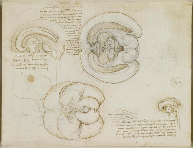
In this sheet Leonardo records an ingenious experiment in which he injected molten wax into the brain to determine the shape of its internal cavities. The drawing at upper centre shows the brain cut in half through the mid-line and opened out; below is a depiction of the base of the brain. The other two studies show the three-dimensional form of the ventricles with reasonable accuracy, the lateral ventricles curving like horns to either side of the third (middle) ventricle, somewhat enlarged and distorted by the pressure of injection.
Leonardo’s earlier studies of the nerve pathways in the head (eg. RCIN 912603) had adopted the traditional belief that the brain contained three bulbous ventricles arranged in a straight line backwards behind the eyes. Even a rudimentary dissection of the brain would have shown him that the brain does indeed contain cavities, but not of this form. However the soft consistency of the unfixed brain and the difficulty of determining the shape of a cavity must have frustrated Leonardo’s attempts to determine the correct form of the ventricles, and so he performed the procedure described here:
"Make two vent-holes in the horns of the greater ventricles and insert melted wax with a syringe, making a hole in the ventricle of memory [the fourth ventricle, at lower right in each drawing]; and through this hole fill the three ventricles of the brain. Then when the wax has set, dissect off the brain and you will see the shape of the ventricles exactly. But first put fine tubes into the vent-holes so that the air which is in these ventricles is blown out and makes room for the wax which enters into the ventricles."
This simple but brilliant technique, adapted from Leonardo’s knowledge of casting bronze sculpture, is the first recorded instance in medical science of injecting a setting medium into a body cavity.
Leonardo probably used a cow’s brain, larger than a human brain and with the foramen magnum (from which the spinal cord issues) more conveniently positioned at the back of the skull. But, as usual, he adjusted the proportions and contours to accord with the shape of a human brain, and the largest drawings thus appear essentially human in form: in the drawing at upper left, the spinal cord is indicated vertically below, rather than horizontally to the rear as it does in most quadrupeds; and the cerebellum (the lobes at centre right of the largest two drawings) is human in position and relative size.The drawing at upper right shows the brain cut in half through the mid-line and opened out, such that the structures mirror one another. Comparison with a human brain sectioned using modern instruments demonstrates the success of Leonardo’s procecure: the only major flaw is the size of the third (middle) ventricle, enlarged and distorted by the pressure of injection in Leonardo’s images; also, the wax did not penetrate to the superior and inferior horns of the curved lateral ventricles, but the results are nevertheless spectacular. The feathery structure spreading out from the centre of the lower drawing, showing the brain from below, is the vascular structure known as the rete mirabile, found in a number of animals (including the cow) but not in humans.
Text adapted from M. Clayton and R. Philo, Leonardo da Vinci: Anatomist, London, 2012Provenance
Bequeathed to Francesco Melzi; from whose heirs purchased by Pompeo Leoni, c.1582-90; Thomas Howard, 14th Earl of Arundel, by 1630; probably acquired by Charles II; Royal Collection by 1690
-
Creator(s)
Acquirer(s)
-
Medium and techniques
Black chalk, pen and ink
Measurements
20.0 x 26.2 cm (sheet of paper)
Object type(s)
Other number(s)


